This quarter I am teaching a histology unit without a microscopy lab. Wait, histology without microscopes… what!?!? I have never experienced a histology course without a lab component and when I first heard this I was very surprised. How do you teach histology without microscopes? What about the concept of magnification? Isn’t operating a microscope a necessary skill? Then I read a couple of journal articles and considered the merits of a purely virtual histology course.
Unless they are involved in a research project requiring optical microscopy or a pathologist analyzing samples, how frequently does the average researcher or medical professional actually use a light microscope? Could time spent practicing optical microscopy be better used learning other skills more relevant to their studies? Maybe career specific workshops or SPSS training? Microscopes are expensive and require upkeep. Could funds instead be used for resources needed in other courses. *cough cough Gross Anatomy cough*
Virtual microscopy is much more efficient for an institution and the students. A college or university could collect a large bank of images that can be updated continuously and won’t deteriorate over time. Online and long distance students are able to fully participate in labs. Slides can be shared rapidly between institutions without risking damaged or lost mail. Instructors can draw on slides to highlight structures without damaging them and students can compare slides of different magnifications or staining techniques side by side.
Histology curriculum often focuses on identifying structures in tissues and relating cell biology to the function of organ systems more than manual lab work. Students could practice reading slides as part of an active learning activity instead. Could microscopy be a workshop or research elective? Training would still be available for students, but only if they are planning to use this skill. This way students genuinely interested in microscopy could receive more individual attention from faculty.
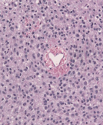
So really, if you consider it, are students losing that much in a histology unit without a microscope? The course I am teaching is “Cell and Tissue Structure and Function.” It is part of the Biochemistry department. Students learn biochemistry and cell biology for the first seven weeks and end with a histology unit from me. We covered the four basic tissue types, integument, circulatory system, cartilage, and bone. The course is part of a physical therapy program. A laboratory component may be important in a course designed for future histologists, but these are physical therapy students. My lectures are packed with images, I have a workshop day set aside to practice analyzing slides, and I think they’ll be okay.
These are some of the papers I read while thinking about this change:
Mione, S., Valcke, M., & Cornelissen, M. (2013). Evaluation of virtual microscopy in medical histology teaching. Anatomical Sciences Education, 6(5), 307-315.
Mione, S., Valcke, M., & Cornelissen, M. (2016). Remote histology learning from static versus dynamic microscopic images. Anatomical Sciences Education, 9(3), 222-230.
Thompson, A. R., & Lowrie, D. J.,Jr. (2017). An evaluation of outcomes following the replacement of traditional histology laboratories with self-study modules. Anatomical Sciences Education, 10(3), 276-285.
Post comes from Julie Doll, MS, Anatomy Instructor in the Department of Anatomy for Chicago College of Osteopathic Medicine at Midwestern University.
I understand (I truly do) the merits of histology w/o microscopes; altho’ as a life-long lover of microscopy, the trend makes me wince. What I WOULD suggest for those who chose to go microscope-less :
(1) Be careful to have enough images so that students are really learning the pattern and not just memorizing a handful of images. So, for example, if they look at enough images of simple squamous epithelium, they should be able to recognize this tissue on a lab practical even in an example that they have never seen before (and that might actually be good practice – you could tell them that they would be tested on images they had never seen, so important to learn the patterns, not memorize the images)
(2) Students already have trouble figuring out what exactly they are looking at when they see a histological specimen in a microscope – that is, there is a disconnect between, say, looking at a slide of liver and seeing a liver in a dissected animal. Something that might help, especially if teaching from images, would be to go the extra mile in showing students how an actual piece of tissue is fixed, embedded, and then sectioned, mounted, and stained. If you can get ahold of some embedded tissues, that might help. At the very least, perhaps show a video of histotechnicians fixing, embedding, and sectioning tissues; to help the students see where the specimen on the slide came from.
My 2 cents.
Though I am “old school” and agree that I am truly wincing as I write this, if the students are truly learning histology then I can see an advantage to both the students and the institution for A&P classes to use digital images instead of teaching students how to use a microscope. I would like to argue that without using the microscope in A&P, students will enter microbiology classes with no prior experience. Unfortunately, despite using the microscope in our A&P classes, the students taking micro after both A&P 1 & 2 normally have little to no proficiency anyway. Further, many have picked up bad habits of using only one eye, not bothering with diopter adjustments, etc.
However, I think there is a disadvantage as well. I think students better appreciate cell size when they see the tissues on the slides before putting them under the microscope. It’s one thing to indicate the magnification of an image. But I think students begin to appreciate how small our cells are when they see nothing more than some purple material on a slide and then suddenly can see individual cells, and how those cells interact to form tissues, as they scan with the microscope. Also, when students hold that tiny letter “e” from newsprint and then see it changing as the image is enlarged, I think they gain more comprehension of magnification, of actual size, and of depth as it will relate to tissues. I’m not sure that students gain that kind of understanding from simply viewing digital images. I think that manually manipulating slides and using the microscope is still an important consideration in A&P.
Perhaps a good approach would be to do both. Begin with the microscope, then progress to digital images. Although, I know that does not help those who want to go microscope-less.