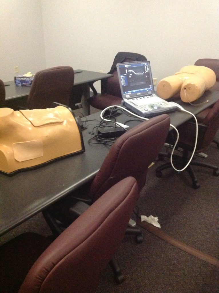
This past weekend was the first Conference on Ultrasound in Human Anatomy and Physiology Education. As President elect of HAPS, I was invited to participate in a panel session during the conference. Not sure of what exactly to expect, I traveled to Columbia, SC for this inaugural conference. I was excited to learn of the possibilities of incorporating ultrasound, but my initial ‘gut’ reaction was that I wouldn’t be able to do too much, since I was not a physician trained in the field. Boy was I wrong!

The first day of the two day conference began with some very informative talks about how various medical schools incorporated ultrasound into their medical school curricula. Among the key points: a) implement in increments (don’t try to set up an entirely new program all at once), b) make sure you assess the students in ultrasound (and don’t just have it as a ‘neato cool toy’ that you never incorporate in exams or other assessments) and c) it isn’t as difficult to use ultrasound as you would think! My response to C initially was “Yeah, right”. I already teach an upper level course entitled Human Anatomy for Medical Imaging, and we do examine ultrasound images in that course. However, I always relied on a skilled ultrasound tech to do ultrasound demonstrations for me, as I had no idea how to even turn on the machine.
Well, boy was I wrong about the difficulty in doing ultrasound demonstrations myself! Don’t get me wrong – being a skilled practitioner of ultrasound takes a LOT of work and training. But I was not aspiring to the level of skilled practitioner. Rather, I became the ‘enthusiastic novice with gross anatomy knowledge’ who was able to pinpoint where major organs were and pick out basic differences between various tissues. With the help of many 1st year medical students from University of South Carolina, I and the other conference participants were able to visualize the common carotid artery and internal jugular vein, determine the difference between the thyroid gland and thyroid cartilage, examine cartilaginous and tendinous structures of the knee joint, visualize the kidneys, spleen, liver, and of course, the heart. Sure, there were times that we were nowhere close to accurately visualizing a particular structure – but with some guidance, we soon learned the basics of the ultrasound machine and some of the tips and tricks to getting a good image. I jumped in and started using the machine on myself – I learned my gallbladder still appears to be ok, my common carotid doesn’t have any major evidence of atherosclerosis, and my creaky right knee still has some cartilage left. 🙂

John Waters (fellow HAPS member) and I quickly thought of possibilities of using ultrasound in the undergraduate A&P classes. It would be very easy to demonstrate key features on the undergraduates and get them excited about visualizing structures in themselves. Whereas prior to the conference, I would not have considered using ultrasound in my intro level human anatomy class, now I was brimming with excitement about the possibilities.
“But what about the cost?” you may ask. That can be a sticking point. Diagnostic-level ultrasound machines can cost 5 or even six digits – well out of range of most undergraduate institutions! But educators in intro classes do not need the ‘best and the brightest’ of ultrasound machines – they need a basic machine that can provide a decent image and is relatively easy to use. Several ultrasound manufacturers are exploring educational partnerships, and are in the process of developing lower-end machines that wouldn’t cost very much for the educator. There may be the possibilities of grant monies to fund these ventures. As local hospitals upgrade their ultrasound equipment, there may be the possibility of your institution being able to purchase the hospital’s older machines. Think outside the box when it comes to funding this venture.
For those of you attending the HAPS conference in Las Vegas this May, you’ll have a chance to see a workshop about incorporating ultrasound in the undergraduate classroom. I hope you will find this concept as interesting as I did!
